Almost all of behavior involves motor function, from talking to gesturing to walking. But even a simple movement like reaching out to pick up a glass of water can be a complex motor task to study. Not only does your brain have to figure out which muscles to contract and in which order to steer your hand to the glass, it also has to estimate the force needed to pick up the glass. Other factors, like how much water is in the glass and what material the glass is made from, also influence the brains calculations. Not surprisingly, there are many anatomical regions which are involved in motor function.
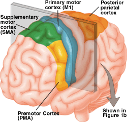
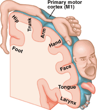
The primary motor cortex, or M1, is one of the principal brain areas involved in motor function. M1 is located in the frontal lobe of the brain, along a bump called the precentral gyrus (figure 1a). The role of the primary motor cortex is to generate neural impulses that control the execution of movement. Signals from M1 cross the bodys midline to activate skeletal muscles on the opposite side of the body, meaning that the left hemisphere of the brain controls the right side of the body, and the right hemisphere controls the left side of the body. Every part of the body is represented in the primary motor cortex, and these representations are arranged somatotopically — the foot is next to the leg which is next to the trunk which is next to the arm and the hand. The amount of brain matter devoted to any particular body part represents the amount of control that the primary motor cortex has over that body part. For example, a lot of cortical space is required to control the complex movements of the hand and fingers, and these body parts have larger representations in M1 than the trunk or legs, whose muscle patterns are relatively simple. This disproportionate map of the body in the motor cortex is called the motor homunculus (figure 1b).


Other regions of the cortex involved in motor function are called the secondary motor cortices. These regions include the posterior parietal cortex, the premotor cortex, and the supplementary motor area (SMA). The posterior parietal cortex is involved in transforming visual information into motor commands. For example, the posterior parietal cortex would be involved in determining how to steer the arm to a glass of water based on where the glass is located in space. The posterior parietal areas send this information on to the premotor cortex and the supplementary motor area. The premotor cortex lies just in front of (anterior to) the primary motor cortex. It is involved in the sensory guidance of movement, and controls the more proximal muscles and trunk muscles of the body. In our example, the premotor cortex would help to orient the body before reaching for the glass of water. The supplementary motor area lies above, or medial to, the premotor area, also in front of the primary motor cortex. It is involved in the planning of complex movements and in coordinating two-handed movements. The supplementary motor area and the premotor regions both send information to the primary motor cortex as well as to brainstem motor regions.
Neurons in M1, SMA and premotor cortex give rise to the fibers of the corticospinal tract. The corticospinal tract is the only direct pathway from the cortex to the spine and is composed of over a million fibers. These fibers descend through the brainstem where the majority of them cross over to the opposite side of the body. After crossing, the fibers continue to descend through the spine, terminating at the appropriate spinal levels. The corticospinal tract is the main pathway for control of voluntary movement in humans. There are other motor pathways which originate from subcortical groups of motor neurons (nuclei). These pathways control posture and balance, coarse movements of the proximal muscles, and coordinate head, neck and eye movements in response to visual targets. Subcortical pathways can modify voluntary movement through interneuronal circuits in the spine and through projections to cortical motor regions.
The spinal cord is comprised of both white and gray matter. The white matter consists of nerve fibers traveling through the spine. It is white because the nerve fibers are insulated with myelin for faster conduction of signals. Like many other large fiber bundles, the corticospinal tract courses through the lateral white matter of the spine. The inside of the spinal cord contains gray matter, composed of the cell bodies of cells including motor neurons and interneurons. In a cross-section of the spinal cord, the shape of the gray matter resembles a butterfly. Fibers in the corticospinal tract synapse onto motor neurons and interneurons in the ventral horn of the spine. Fibers coming from hand regions in the cortex end on motor neurons higher up in the spine (in the cervical levels) than fibers from the leg regions which terminate in the lumbar levels. The lower levels of the spine therefore have much less white matter than the higher levels.
Within the ventral horn, motor neurons projecting to distal muscles are located more laterally than neurons controlling the proximal muscles. Neurons projecting to the trunk muscles are located the most medially. Furthermore, neurons of extensors (muscles that increase the joint angle such as the triceps muscle) are found near the edge of the gray matter, but the flexors (muscles which decrease the joint angle such as the biceps muscle) are more interior. It is important to note that a single motor neuron in the spine can receive thousands of inputs from the cortical motor regions, the subcortical motor regions and also through interneurons in the spine. These interneurons receive input from the same regions, and allow complex circuits to develop.
Signals generated in the primary motor cortex travel down the corticospinal tract (green) through the spinal white matter to synapse on interneurons and motor neurons in the spinal cords ventral horn. Ventral horn neurons in turn send their axons (blue) out through the ventral roots to innervate individual muscle fibers. In this example, a signal from M1 travels through the corticospinal tract and exits the spine around the sixth cervical level. A peripheral motor neuron relays the signal out to the arm to activate a group of myofibrils in the bicep, causing that muscle to contract. Collectively, the ventral horn motor neuron, its axon, and the myofibrils that it innervates are called a single motor unit.
Each motor neuron in the spine is part of a functional unit called the motor unit (figure 2). The motor unit is composed of the motor neuron, its axon and the muscle fibers it innervates. Smaller motor neurons typically innervate smaller muscle fibers. Motor neurons can innervate any number of muscle fibers, but each fiber is only innervated by one motor neuron. When the motor neuron fires, all of its muscle fibers contract. The size of the motor units and the number of fibers that are innervated contribute to the force of the muscle contraction.
There are two types of motor neurons in the spine, alpha and gamma motor neurons. The alpha motor neurons innervate muscle fibers that contribute to force production. The gamma motor neurons innervate fibers within the muscle spindle. The muscle spindle is a structure inside the muscle that measures the length, or stretch, of the muscle. The role of the musclespindle in reflexes such as the knee jerk reflex will be reviewed in the Motor Systems Physiology section of this NeuroSeries. The golgi tendon organ is also a stretch receptor, but it is located in the tendons that connect the muscle to the skeleton. It provides information to the motor centers about the force of the muscle contraction. Information from muscle spindles, golgi tendon organs and other sensory organs are directed to the cerebellum. The cerebellum is a small grooved structure located in the back of the brain beneath the occipital lobe. This motor region is specifically involved when learning a new sport or dance step or instrument. The cerebellum is involved in the timing and coordination of motor programs. The actual motor programs are generated in the basal ganglia. The basal ganglia are several subcortical regions that are involved in organizing motor programs for complex movements. Damage to these regions result in spontaneous, inappropriate movements. The basal ganglia send output to other subcortical brain regions and the cortex.
Through the interaction of many anatomical motor regions, everyday movements seem effortless and more complex movements can be learned.


